• Skip to primary navigation
  • Skip to main content

The Digital Pathology Company

  • ‹ Company website
  • For Partner
  • Software Downloads
  • Old navigation ›
    • Homepage
    • Solutions
      • Solutions for Diagnostics
      • Solutions for Research
      • Solutions for Education
      • Solutions for Veterinary Pathology
      • Solutions for Geology
      • Scanning Service
    • Diagnostics
      • PANNORAMIC Pathology Management System
        • CaseManager DX
          • ClinicalViewer
        • DiagnosticApplications
      • PANNORAMIC Diagnostic Scanners
        • PANNORAMIC Flash DESK DX
        • Digital Pathology Solution with The Pannoramic 75 DX Digital Scanner
        • Digital Pathology Solution with The Pannoramic 150 DX Digital Scanner
        • PANNORAMIC 250 Flash III DX
        • PANNORAMIC™ 480 DX
        • PANNORAMIC 1000 DX
        • Legacy Scanners
      • Laboratory Information System
        • Track & Sign
        • CentralLogService Application (CLS) – Cybersecurity
      • Laboratory Components
        • MacroStation
        • SlideDriver
    • Research
      • SlideManager
      • PANNORAMIC™ Digital Slide Scanners
        • PANNORAMIC 1000
        • PANNORAMIC™ 480
        • PANNORAMIC 250 Flash III
        • PANNORAMIC Confocal
        • PANNORAMIC SCAN II
        • PANNORAMIC MIDI II
        • Pannoramic™ MIDI III
        • PANNORAMIC DESK II DW
      • Tissue Microarrayers
        • TMA Grand Master
        • TMA molmed
        • TMA Master II
        • TMA Applications
          • TMA Control
          • TMA Module
      • PANNORAMIC-X
      • Digital Microscopes – Viewers
        • SlideViewer
        • WebViewer
        • InstantViewer
        • iPad Viewer
        • 3DView
      • Image Analysis – QuantCenter
      • Slide Management – SlideCenter
        • SlideMaster
      • SimpleSlideInterface
      • ESchool
      • Software Downloads
    • Sales
      • Newsletter
      • Inquiry
      • Distributor Network
      • Become a Distributor
    • Support
      • Technical Support Form
      • Activity Report Form for Partners
      • Knowledge Base
        • Reference Institutes
        • Publications
          • Publications
          • TMA Grand Master publication list
          • TMA Master publication list
        • Case Studies
        • Whitepapers
        • Tutorials
      • Request Demo
    • News & Events
      • Events
      • News
    • About Us
      • Awards
      • Quality Management System
      • Funds
      • Funds (HU)
      • Contact Us
    • Feedback
  • Partner Home
  • Account
  • Log Out
Home / Digital TMA Workflow

Digital TMA Workflow

The tissue microarray (TMA) technique brings revolutionary changes in high-throughput diagnosis by being able to handle up to several hundred samples in one block or in one slide. With the TMA technique you can save a considerable amount of time, reagents, and slide storage while achieving more standardized laboratory conditions. Tissue microarrays can make working with and evaluation of IHC, FISH and other staining protocols faster and easier.

3DHISTECH’s tissue microarray product range covers every step of the digital TMA workflow.

TMA WORKFLOW

Step 1  

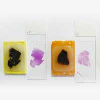
Sample designation and slide preparation from the donor blocks

Step 2  

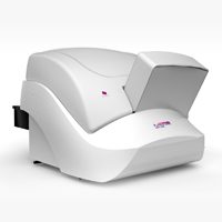
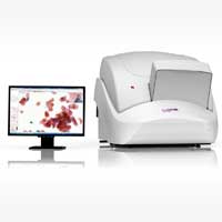
Slide scanning with a Pannoramic digital slide scanner
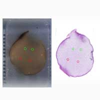
Step 3  

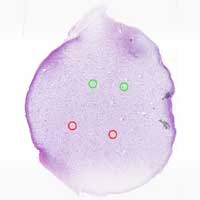
Marking the representative tissue regions

Step 4  

Block layout design in tha TMA Grand Master software

Step 5 

Digital slide overlay and marker transfer
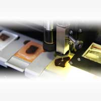
Step  

Punching from donor block into recipient block and saving of Excel database

Step 7  

TMA slide scanning with a Pannoramic digital slide scanner
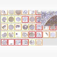
Step 8

Evaluation of the TMA project using the TMA software and the Excel database

You need

Standardized solutions
For immunohistochemistry, constant quality is essential in all workflow phases. Whether it is tissue microarray block building, slide scanning or digital image analysis, you can be sure 3DHISTECH products provide the best quality.

High quality brightfield scanning
The Pannoramic SCAN digital slide scanner won the 1st prize of ‘Quality Scan 40x’ category at the first European Scanner Contest in 2010. The same optical path is used in all of the other Pannoramic scanners, delivering the best quality images to your screen in minutes!

Automated image quantification tools
Advanced image analysis algorithms are proved to be helpful in diagnosis of IHC stained samples. 3DHISTECH’s automated IHC quantification software are robust, powerful and cope with different stain intensities.

Workflow tools
More stains mean more data and you need to channel this huge amount of information into easily managable projects. The immunohistochemistry and TMA workflow tools from 3DHISTECH offer a flexible and standardized way to deal with these challenges.

Reviewing and reporting tools
The final step in the pathology workflow is of vital importance. You need to present your diagnosis data in an easy-to-understand and pleasing way. With the 3DHISTECH reviewing and reporting tools you can achieve this faster.

Product certificate

Product brochure

  • ‹ Company website
  • For Partners
  • Software Downloads
  • facebook
  • X
  • linkedin
  • youtube
  • instagram

Partners

Login

© 2005-2025, 3DHISTECH Ltd. All rights reserved.
H-1141 Budapest, Öv u. 3., Hungary

We are using cookies to give you the best experience on our website.

You can find out more about which cookies we are using or switch them off in .

3DHISTECH Ltd.
Powered by  GDPR Cookie Compliance
Privacy Overview

This website uses cookies so that we can provide you with the best user experience possible. Cookie information is stored in your browser and performs functions such as recognising you when you return to our website and helping our team to understand which sections of the website you find most interesting and useful.

Strictly Necessary Cookies

Strictly Necessary Cookie should be enabled at all times so that we can save your preferences for cookie settings.

If you disable this cookie, we will not be able to save your preferences. This means that every time you visit this website you will need to enable or disable cookies again.

Google Analytics

This website uses Google Analytics to collect anonymous information such as the number of visitors to the site, and the most popular pages.

Keeping this cookie enabled helps us to improve our website.

Please enable Strictly Necessary Cookies first so that we can save your preferences!