Please, visit our Booth#B38 at MEDLAB Asia Pacific taking place at the Marina Bay Sands, where we present our brand new products, applications and highlights.
Date: 18th – 20th of February, 2014
Location: Singapore, Malaysia
Please, visit our Booth#B38 at MEDLAB Asia Pacific taking place at the Marina Bay Sands, where we present our brand new products, applications and highlights.
Date: 18th – 20th of February, 2014
Location: Singapore, Malaysia
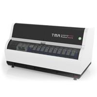
The tissue microarray (TMA) technique can be used as a valuable, high-throughput diagnostic method. By being able to place up to several hundred different samples into one paraffin block, TMA brings major economies in time quality and costs of tissue preparation, staining and slide preparation. The real advantages of tissue microarrays can only be achieved when using digital TMA. With 6 PCR cassettes and 10 PCR tubes / cassette.
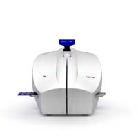
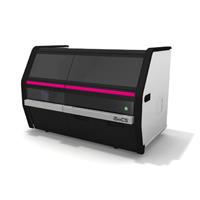
The Pannoramic MIDI II is an automatic digital slide scanner with a remarkable feature set at an affordable price: 12-slide capacity, fluorescence scanning, and many more. Reliable digital slide scanner for the medium-sized laboratory. Its superior image quality and features in brightfield and fluorescence make it the right choice for research purposes.
• 12 slide capacity
• Brightfield and 9-channel fluorescent scanning
• Wet slide compatible
• Manual camera changer
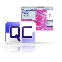
QuantCenter is the framework for 3DHISTECH image analysis applications. By first segmenting the sample on the tissue level, the cell based algorithms can run faster and provide more reliable results. QuantCenter is designed for predictive and prognostic marker quantification and can be adapted to your lab’s IHC protocols.
Teach your software to detect the areas of interest on your slides automatically! Run quantification algorithms on automatically detected ROIs to save time and effort!
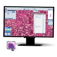
The new NuclearQuant software application performs automatic evaluation on IHC nuclear stainings (ER, PR, Ki67, etc.). Using color deconvolution it measures staining intensity only on the chromogen channel. The algorithm categorizes the detected nuclei to negative, weak positive, medium positive and strong positive classes. The new classification gallery is also available for locating the detections one by one and rescoring.
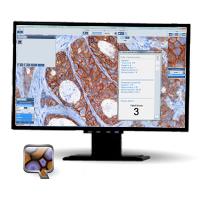
The new MembraneQuant software application offers automated evaluation for IHC membrane stainings (HER-2, EGFR, etc.). With the help of color deconvolution the intensity measurements are performed only on the chromogen channel. The algorithm categorizes the detected cells (+, ++, +++), and determines the H-score for the annotated area. In the classification gallery can view, find and also rescore the detected membranes.
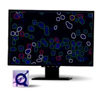
In the 1.6.2 release we made the measurement possibilities of FISHQuant complete. Not only can FISHQuant work on cell samples, but the image processing of tissue samples is also possible. The new program, while maintaining the functionality of Cyto FISH, received a secondary non-specific measurement option, which allows for the numerical analysis of translocation probes, containing no structural abnormalities, and display the combined results of the two measurements and statistical analysis.
Brand new application software implementing two different algorithms, CISH and CISH-RNA to the chromomeric in situ hybridization patterns, perfectly suited for detecting viral RNA.
Brand new application software implementing two different algorithms, CISH and CISH-RNA to the chromomeric in situ hybridization patterns, perfectly suited for detecting viral RNA
Upcoming conferences where 3DHISTECH will be present:
USCAP
3 – 5 March 2014
San Diego, CA, USA
Termine und Demonstrationen in Österreich
14 – 15 March 2014
Wien, Austria
AACR
6 – 9 April 2014
San Diego, CA, USA
Pathology Informatics Summit
13 – 16 May 2014
Pittsburg, PA, USA
III. Pannonia Congress of Pathology
15 – 17 May 2014
Bled, Slovenia