• Skip to primary navigation
  • Skip to main content

The Digital Pathology Company

  • ‹ Company website
  • For Partner
  • Software Downloads
  • Old navigation ›
    • Homepage
    • Solutions
      • Solutions for Diagnostics
      • Solutions for Research
      • Solutions for Education
      • Solutions for Veterinary Pathology
      • Solutions for Geology
      • Scanning Service
    • Diagnostics
      • PANNORAMIC Pathology Management System
        • CaseManager DX
          • ClinicalViewer
        • DiagnosticApplications
      • PANNORAMIC Diagnostic Scanners
        • PANNORAMIC Flash DESK DX
        • Digital Pathology Solution with The Pannoramic 75 DX Digital Scanner
        • Digital Pathology Solution with The Pannoramic 150 DX Digital Scanner
        • PANNORAMIC 250 Flash III DX
        • PANNORAMIC™ 480 DX
        • PANNORAMIC 1000 DX
        • Legacy Scanners
      • Laboratory Information System
        • Track & Sign
        • CentralLogService Application (CLS) – Cybersecurity
      • Laboratory Components
        • MacroStation
        • SlideDriver
    • Research
      • SlideManager
      • PANNORAMIC™ Digital Slide Scanners
        • PANNORAMIC 1000
        • PANNORAMIC™ 480
        • PANNORAMIC 250 Flash III
        • PANNORAMIC Confocal
        • PANNORAMIC SCAN II
        • PANNORAMIC MIDI II
        • Pannoramic™ MIDI III
        • PANNORAMIC DESK II DW
      • Tissue Microarrayers
        • TMA Grand Master
        • TMA molmed
        • TMA Master II
        • TMA Applications
          • TMA Control
          • TMA Module
      • PANNORAMIC-X
      • Digital Microscopes – Viewers
        • SlideViewer
        • WebViewer
        • InstantViewer
        • iPad Viewer
        • 3DView
      • Image Analysis – QuantCenter
      • Slide Management – SlideCenter
        • SlideMaster
      • SimpleSlideInterface
      • ESchool
      • Software Downloads
    • Sales
      • Newsletter
      • Inquiry
      • Distributor Network
      • Become a Distributor
    • Support
      • Technical Support Form
      • Activity Report Form for Partners
      • Knowledge Base
        • Reference Institutes
        • Publications
          • Publications
          • TMA Grand Master publication list
          • TMA Master publication list
        • Case Studies
        • Whitepapers
        • Tutorials
      • Request Demo
    • News & Events
      • Events
      • News
    • About Us
      • Awards
      • Quality Management System
      • Funds
      • Funds (HU)
      • Contact Us
    • Feedback
  • Partner Home
  • Account
  • Log Out
Home / News / Invitation to attend the ECP 2014

Invitation to attend the ECP 2014

2019.09.20. by

European Congress of Pathology 2014

3DHISTECH cordially invites you to attend the 26th European Congress of Pathology
(ECP 2014, http://www.esp-congress.org)
Date: 30th of August – 3rd of September, 2014
Location: ExCeL London; One Western Gateway, Royal Victoria Dock, London E16 1XL, UK

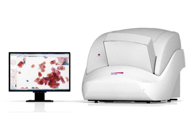
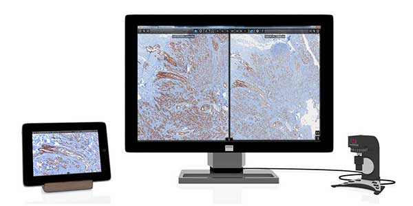
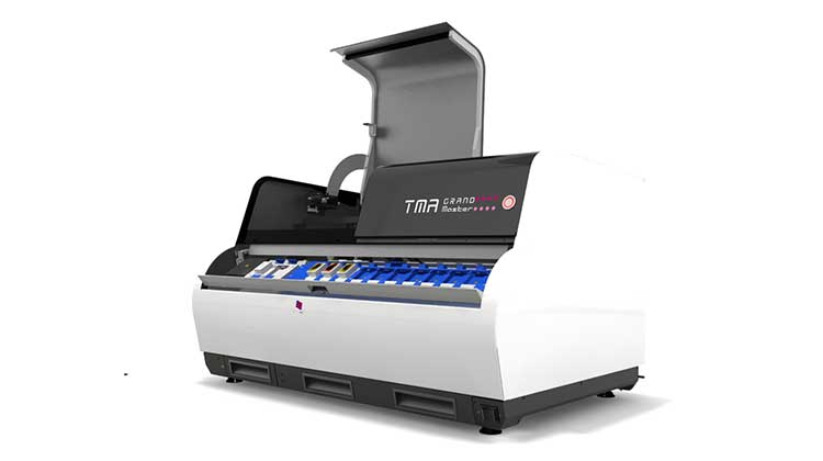
3DHISTECH is the Major Sponsor of ECP 2014 and will exhibit at booth #7 in the main Exhibition Hall.

3DHISTECH invites you to attend its Luncheon Symposium during the ECP 2014.
Title of the symposium: “Digitalisation in routine and research pathology.”
Date: Monday, September 1, 2014
Time: 13.00-14.30
Room: Capital Suite 8/11

Guest speakers and their presentations:
Dr. Asel Kudaybergenova
Pathologist; Russian Science Center of Radiology and Surgical Technology (RSCRST), RUSSIA
Presentation title: 
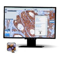
“Quantitative measurements of cell density and proliferative markers by image analysis as approach to make the pathology report more objective.”

Prof. Ince Ümit, MD
Chief Pathologist; Acibadem Health Group Pathology Dept., TURKEY
Presentation title: “Validated Interhospital Routine Digital Pathology / Telepathology Practice in a Multi-institutional Health Group in Turkey.”

Dr. Peter March
Bioimaging Senior Experimental Officer; The University of Manchester, UK
Presentation title: “The use of digital whole slide scanning in academic research. The power of having everything warts and all!”

Dr. Yevgeniy Romin, MS
Digital Microscopy Specialist; Memorial Sloan Kettering Cancer Center, New York, USA
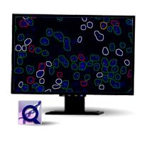
Presentation title: “Quantification of brightfield and multi-color fluorescent signal in normal tissues and in tumors using digitally scanned serial sections.”

Featuring

Pannoramic 250 Flash II

According to the results of the 2nd International Scanner Contest (ISC), this is the fastest digital slide scanner on the market at both 20x and 40x resolution. Designed for routine digital pathology, the new Flash II easily meets your needs, from high quality to high capacity.

Digital Pathology Cockpit Advanced

3DHISTECH is introducing the Digital Pathology Cockpit which combines the comfort of microscope navigation with the highest quality digital slides. What is more, the Digital Pathology Cockpit allows you to fully utilize the advantages of digital slides in the clinical environment.

TMA Grand Master

TMA Grand Master with PCR extraction function. The tissue microarray (TMA) technique can be used as a valuable, high-throughput diagnostic method. By being able to place up to several hundred different samples into one paraffin block, TMA brings major economies in time quality and costs of tissue preparation, staining and slide preparation. The real advantages of tissue microarrays can only be achieved when using digital TMA. With 6 PCR cassettes and 10 PCR tubes/cassette.

Pannoramic Confocal

3DHISTECH has managed to couple confocal imaging with the Pannoramic award-winning whole slide scanning technology. With Pannoramic Confocal your immunofluorescent samples will appear on your monitor in unprecedented quality.
• 6 channel fluorescence illumination with solid state light source
• Highest confocal light efficiency
• Brightfield overlay

CaseCenter

Always be closer than three clicks from any of your slides. CaseCenter is a robust web-based database for storing, managing and distributing your slides. VirtualTray collects slides and images of a whole case into one continuous view that makes your daily work more efficient than ever before.

Viewers

Speed up your work and meet the most ergonomic and sate-of-the art WSI viewer software. CaseViewer is designed for the routine daily work of pathologists using whole slide images instead of a regular microscope. Supports new VirtualTray feature for the highest efficiency.

QuantCenter

QuantCenter is the framework for 3DHISTECH image analysis applications. By first segmenting the sample on the tissue level, the cell based algorithms can run faster and provide more reliable results. QuantCenter is designed for predictive and prognostic marker quantification and can be adapted to your lab’s IHC protocols.

PatternQuant

Teach your software to detect the areas of interest on your slides automatically! Run quantification algorithms on automatically detected ROIs to save time and effort!

NuclearQuant

The new NuclearQuant software application performs automatic evaluation on IHC nuclear stainings (ER, PR, Ki67, etc.). Using color deconvolution it measures staining intensity only on the chromogen channel. The algorithm categorizes the detected nuclei to negative, weak positive, medium positive and strong positive classes. The new classification gallery is also available for locating the detections one by one and rescoring.

MembraneQuant

The new MembraneQuant software application offers automated evaluation for IHC membrane stainings (HER-2, EGFR, etc.). With the help of color deconvolution the intensity measurements are performed only on the chromogen channel. The algorithm categorizes the detected cells (+, ++, +++), and determines the H-score for the annotated area. In the classification gallery can view, find and also rescore the detected membranes.

FISHQuant

In the 1.6.2 release we made the measurement possibilities of FISHQuant complete. Not only can FISHQuant work on cell samples, but the image processing of tissue samples is also possible. The new program, while maintaining the functionality of Cyto FISH, received a secondary non-specific measurement option, which allows for the numerical analysis of translocation probes, containing no structural abnormalities, and display the combined results of the two measurements and statistical analysis.

CISHQuant

Brand new application software implementing two different algorithms, CISH and CISH-RNA to the chromomeric in situ hybridization patterns, perfectly suited for detecting viral RNA.

3DView

CT for microscopy! 3DView can show you the reconstructed model of the tissue in microscopic details.
• Works with digital slides or standard images of serial sections
• Automatic alignment of consecutive layers
• Flexible visualization options (sectioning plane, HE masking, separate FL channel management, etc.)
• 2D and volumetric measurements

  • ‹ Company website
  • For Partners
  • Software Downloads
  • facebook
  • X
  • linkedin
  • youtube
  • instagram

Partners

Login

© 2005-2025, 3DHISTECH Ltd. All rights reserved.
H-1141 Budapest, Öv u. 3., Hungary

We are using cookies to give you the best experience on our website.

You can find out more about which cookies we are using or switch them off in .

3DHISTECH Ltd.
Powered by  GDPR Cookie Compliance
Privacy Overview

This website uses cookies so that we can provide you with the best user experience possible. Cookie information is stored in your browser and performs functions such as recognising you when you return to our website and helping our team to understand which sections of the website you find most interesting and useful.

Strictly Necessary Cookies

Strictly Necessary Cookie should be enabled at all times so that we can save your preferences for cookie settings.

If you disable this cookie, we will not be able to save your preferences. This means that every time you visit this website you will need to enable or disable cookies again.

Google Analytics

This website uses Google Analytics to collect anonymous information such as the number of visitors to the site, and the most popular pages.

Keeping this cookie enabled helps us to improve our website.

Please enable Strictly Necessary Cookies first so that we can save your preferences!