• Skip to primary navigation
  • Skip to main content

The Digital Pathology Company

  • ‹ Company website
  • For Partner
  • Software Downloads
  • Old navigation ›
    • Homepage
    • Solutions
      • Solutions for Diagnostics
      • Solutions for Research
      • Solutions for Education
      • Solutions for Veterinary Pathology
      • Solutions for Geology
      • Scanning Service
    • Diagnostics
      • PANNORAMIC Pathology Management System
        • CaseManager DX
          • ClinicalViewer
        • DiagnosticApplications
      • PANNORAMIC Diagnostic Scanners
        • PANNORAMIC Flash DESK DX
        • Digital Pathology Solution with The Pannoramic 75 DX Digital Scanner
        • Digital Pathology Solution with The Pannoramic 150 DX Digital Scanner
        • PANNORAMIC 250 Flash III DX
        • PANNORAMIC™ 480 DX
        • PANNORAMIC 1000 DX
        • Legacy Scanners
      • Laboratory Information System
        • Track & Sign
        • CentralLogService Application (CLS) – Cybersecurity
      • Laboratory Components
        • MacroStation
        • SlideDriver
    • Research
      • SlideManager
      • PANNORAMIC™ Digital Slide Scanners
        • PANNORAMIC 1000
        • PANNORAMIC™ 480
        • PANNORAMIC 250 Flash III
        • PANNORAMIC Confocal
        • PANNORAMIC SCAN II
        • PANNORAMIC MIDI II
        • Pannoramic™ MIDI III
        • PANNORAMIC DESK II DW
      • Tissue Microarrayers
        • TMA Grand Master
        • TMA molmed
        • TMA Master II
        • TMA Applications
          • TMA Control
          • TMA Module
      • PANNORAMIC-X
      • Digital Microscopes – Viewers
        • SlideViewer
        • WebViewer
        • InstantViewer
        • iPad Viewer
        • 3DView
      • Image Analysis – QuantCenter
      • Slide Management – SlideCenter
        • SlideMaster
      • SimpleSlideInterface
      • ESchool
      • Software Downloads
    • Sales
      • Newsletter
      • Inquiry
      • Distributor Network
      • Become a Distributor
    • Support
      • Technical Support Form
      • Activity Report Form for Partners
      • Knowledge Base
        • Reference Institutes
        • Publications
          • Publications
          • TMA Grand Master publication list
          • TMA Master publication list
        • Case Studies
        • Whitepapers
        • Tutorials
      • Request Demo
    • News & Events
      • Events
      • News
    • About Us
      • Awards
      • Quality Management System
      • Funds
      • Funds (HU)
      • Contact Us
    • Feedback
  • Partner Home
  • Account
  • Log Out
Home / Research / Digital Microscopes – Viewers / 3DView

3DView

  • microCT image in 3DView

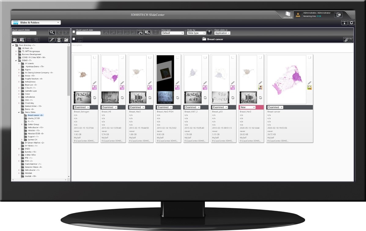
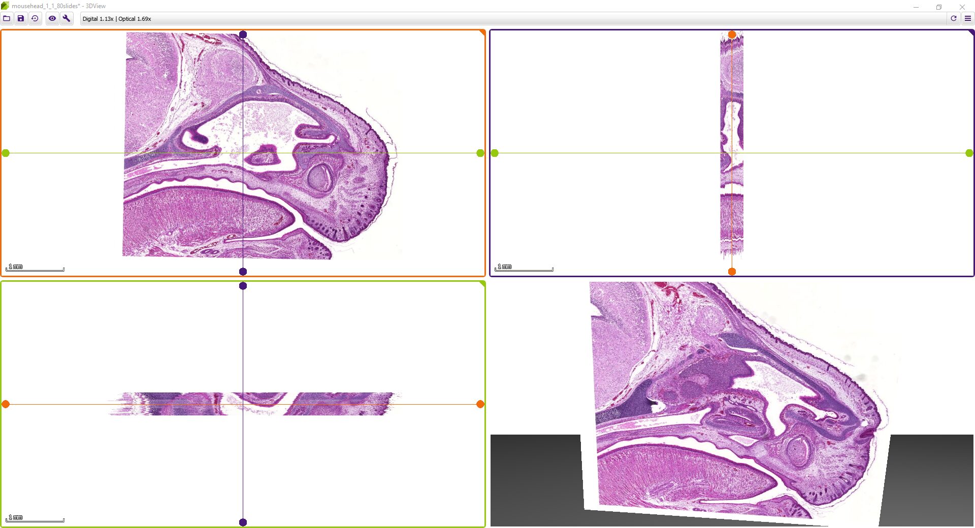
3DView by 3DHISTECH is a software application that prepares 3D reconstructions of 2D serial sections. It is also capable of displaying microCT images in 3D (VOL/VGI format).

While microscope slides allow you to see only one section of reality, 3DView is a tool that can reconstruct the original tissue from its serial sections. The 3D image can then be rotated, zoomed into and “sliced” according to user needs.

How 3DView Works

  • Aligns several 2D serial sections using the SlideMatch function;
  • Prepares a 3D reconstruction from 2D serial sections;
  • The 3D image can then be rotated, zoomed into and “sliced” according to user needs.

Key features

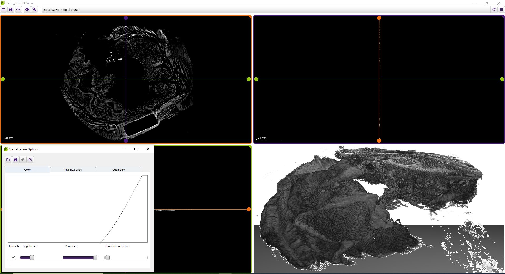
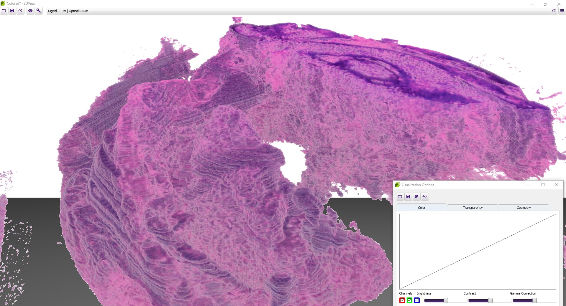
Advanced visualization options

  • Besides brightness, contrast, gamma, RGB channels, etc., the user can select and edit several stainings for best visualization.
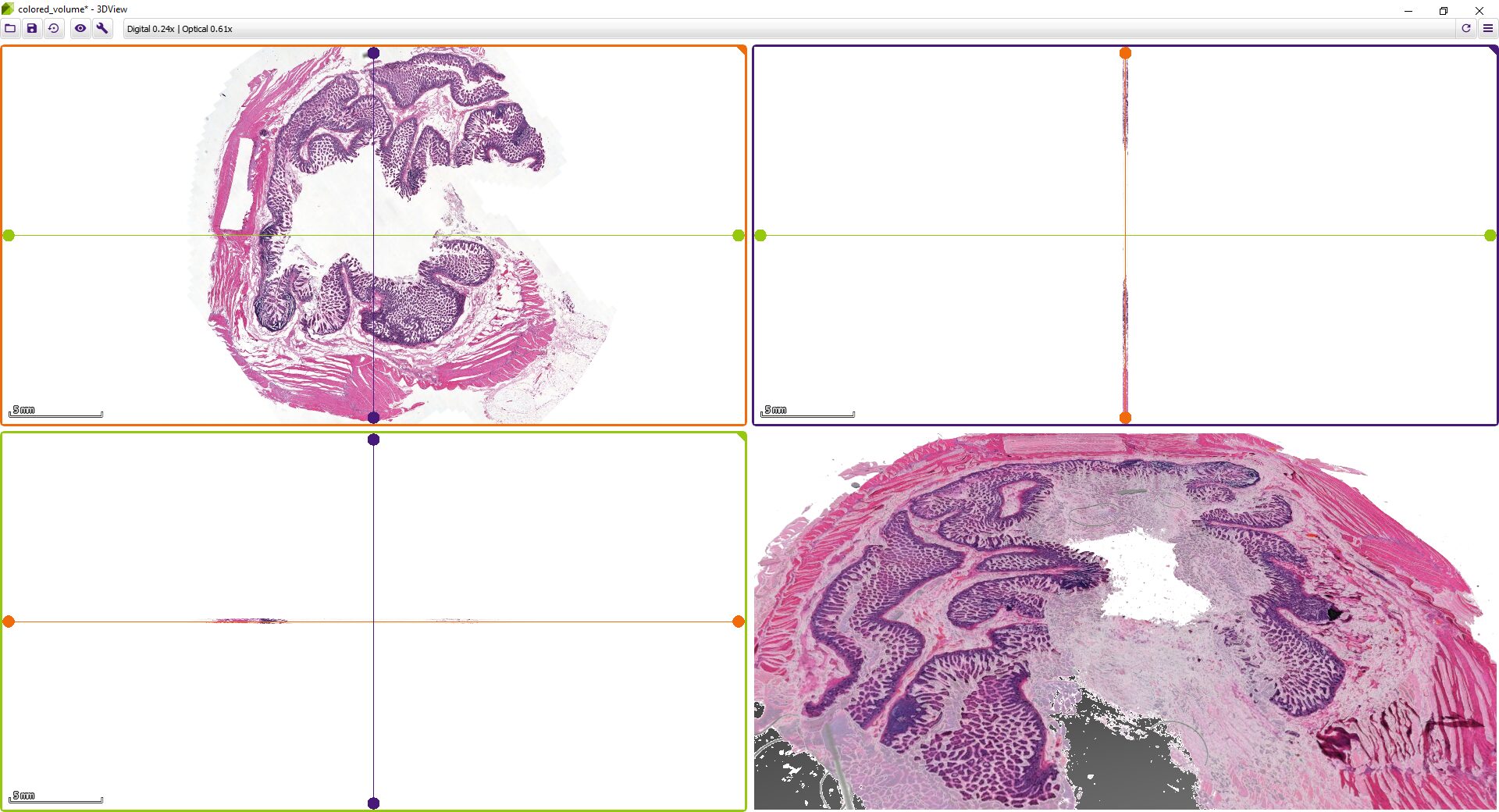
  • Slice view (2D) of arbitrary sections and volume view (3D) of a volumetric data set are both available.
  • 3DView is also capable of preparing 3D reconstructions from a Z-stack slide.

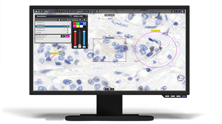
Measurement tools to assess metric distances

Advanced file export options

  • The reconstruction can be exported as either 2D series or a 3D volume, with several channel data, alignment, export magnification and file format options – including high-resolution video.
  • High-resolution snapshot export for publications is also available.

Wide compatibility with third-party file formats

Besides MRXS by 3DHISTECH, 3DView handles SVS by Leica Aperio, CZI by Zeiss, NDP by Hamamatsu, and VSI by Olympus.

3dview on a monitor

Minimum system requirements

  • Operating system: Windows 7 SP1, 8. 8.1 or 10 (64-bit version)
  • Graphics processing unit: OpenGL 3.3 capable (with dedicated video memory of 512 MB minimum, 1 GB recommended), such as from AMD/ATI, Barco, Intel, or NVIDIA
  • CPU: Intel Core i7 or comparable
  • RAM: 4 GB minimum, 8 GB recommended

Would you like to know more about our solutions? Please get in touch!

Related Products

SlideCenter

SlideCenter (former CaseCenter) is a powerful, server-based slide management system with a slide database.
slide management

iPad Viewer

Experience true mobile pathology!
iPad Viewer

InstantViewer

Open and share your slides with the multi-platform InstantViewer!
InstantViewer by3DHISTECH

  • ‹ Company website
  • For Partners
  • Software Downloads
  • facebook
  • X
  • linkedin
  • youtube
  • instagram

Partners

Login

© 2005-2025, 3DHISTECH Ltd. All rights reserved.
H-1141 Budapest, Öv u. 3., Hungary

We are using cookies to give you the best experience on our website.

You can find out more about which cookies we are using or switch them off in .

3DHISTECH Ltd.
Powered by  GDPR Cookie Compliance
Privacy Overview

This website uses cookies so that we can provide you with the best user experience possible. Cookie information is stored in your browser and performs functions such as recognising you when you return to our website and helping our team to understand which sections of the website you find most interesting and useful.

Strictly Necessary Cookies

Strictly Necessary Cookie should be enabled at all times so that we can save your preferences for cookie settings.

If you disable this cookie, we will not be able to save your preferences. This means that every time you visit this website you will need to enable or disable cookies again.

Google Analytics

This website uses Google Analytics to collect anonymous information such as the number of visitors to the site, and the most popular pages.

Keeping this cookie enabled helps us to improve our website.

Please enable Strictly Necessary Cookies first so that we can save your preferences!