SlideViewer (formerly CaseViewer) is a digital microscopy application designed for supporting the microscope examination process in bioscience.
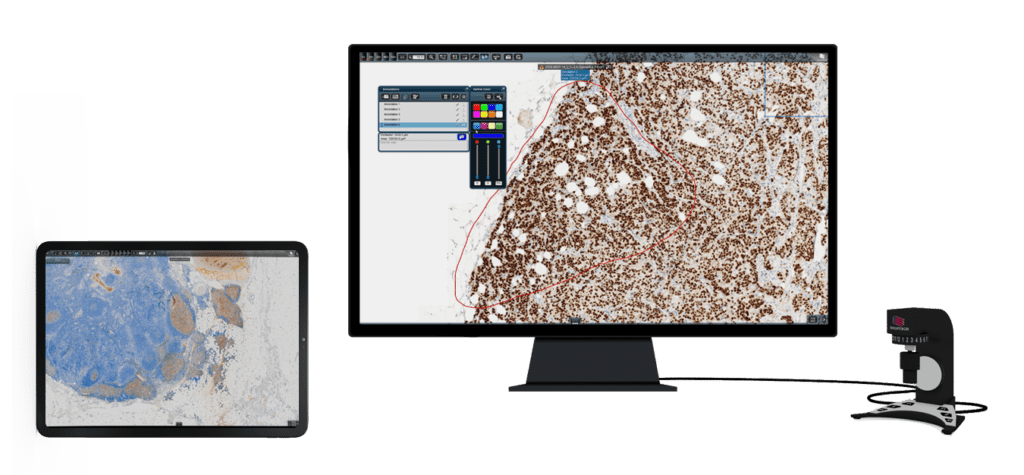
Researchers and medical students need user-friendly digital microscopy tools for the purposes of research and study that can be used across a variety of platforms and devices.
SlideViewer, 3DHISTECH’s advanced slide viewing software provides researchers, lecturers and students with a flexible and easy-to-use digital microscopy tool for more accurate research results and state-of-the-art pathology education.

Main New Features (SlideViewer 2.8)
New Z-Stack control interface
- Redesigned stepping, auto-stepping and prefetch buttons.
Settings menu / Playback option for Z-stack layers
It is possible to set in Settings menu how the Z-stack layers are played:
- stop at the end,
- or playback continues continuously when the end is reversed,
- or restart from the beginning at the end and play continuously.
Switching between Z-Stack layers with SlideDriver
- It is possible to switch between Z-layers with SlideDriver, if the Z-stack slide is open and the Z-stack is on, the big dial will switch Z-layers.
3DConnexion mouse support – 3DConnexion Settings
The Settings menu has a new 3DConnexion tab with the following options:
- Motion: Zoom dependent, Fixed
- Speed (a slider to set the speed of X/Y navigation)
- Inversion option: X, Y, and zoom.
Savable color setting per staining
- It is possible to assign a color setting to the stain type and save and apply it to other stained slides opened from CaseCenter/SlideCenter, if the stain information is filled in.
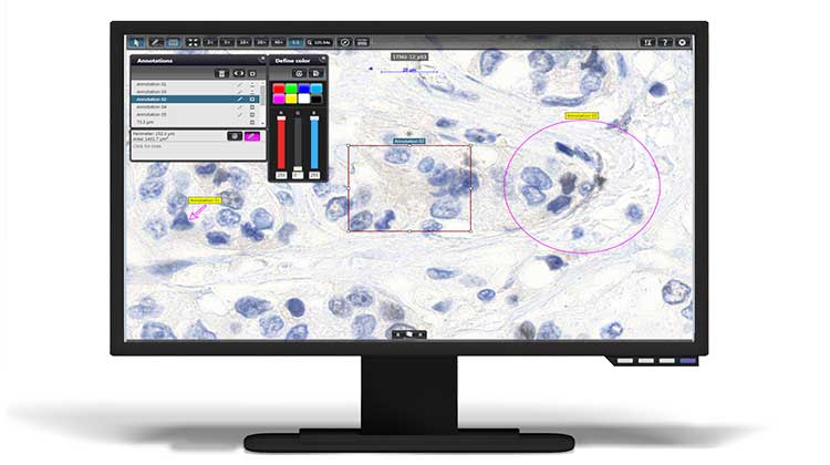
Annotation shape changing
- The shape of a finished annotation can be modified after selection. It is possible to bring up the editor with double click on the selected open or closed polygon annotation and modify its shape.
Displaying the scanned area on the whole slide preview
- The ‘Whole slide preview’ of the selected slide, the scanned area is highlighted in red.
Minimum System Requirements
- Monitor resolution 1280 × 720
- Operation system Windows 8.1 or newer
- Graphics Card OpenGL 2.0 compatible
- CPU Intel DualCore
- RAM 2GB
New Features
Easy slide viewing and navigation
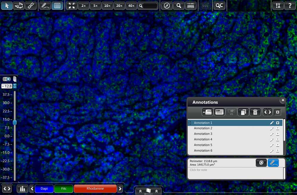
- Ability to search for annotation name, note and user.
- A grid can be added to the opened slide for analysis. The properties of the grid can be customized in the settings menu.
- The annotation labels now displays extra information.
- Possibility to display digital slides and their colors based on previously saved histogram profiles.
- Possibility to change to a gray-scale mode.
- Usability improvements – Touch friendly settings panel and 3DConnexion SpaceMouse control. Ability to jump one screen size from chosen field of view. Displays the name of the file type on the top of the UI. Capable of automatic slide rotation in preview mode. Navigate between layers in Z-Stack.
Key Features (General)
Easy slide viewing and navigation
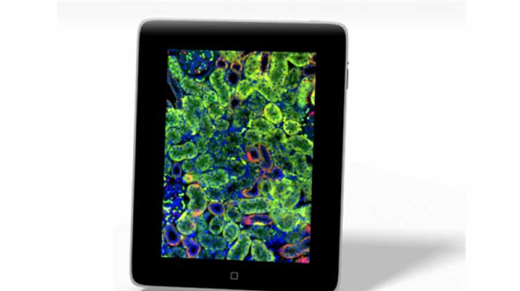
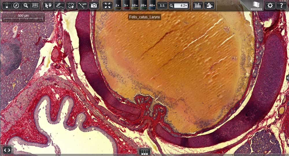
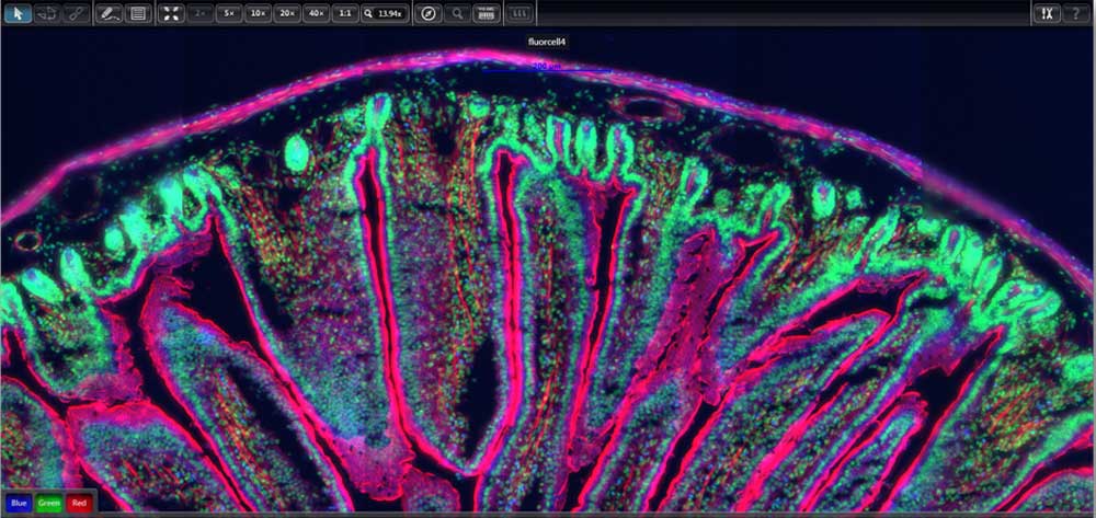
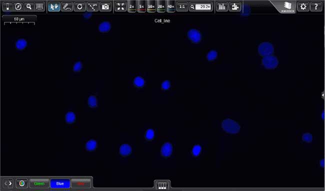
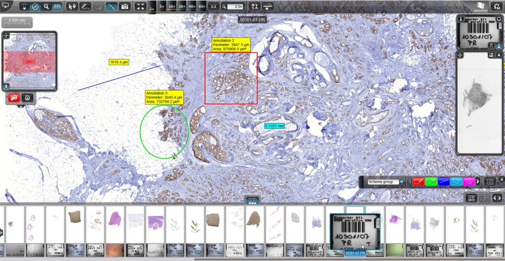
- A simple and straightforward graphical user interface, suited for viewing brightfield, fluorescence and confocal slides.
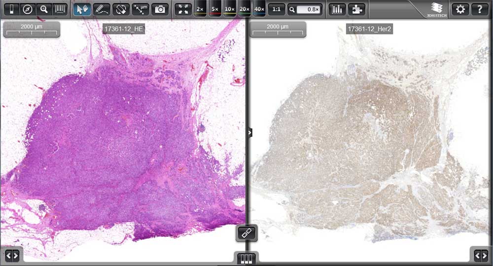
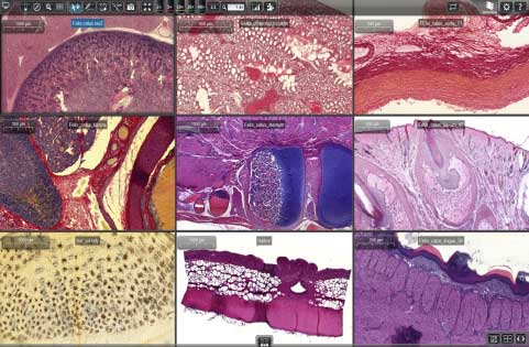
- MultiView: A maximum of nine slides of a single case with different stainings can be viewed simultaneously on the main screen.
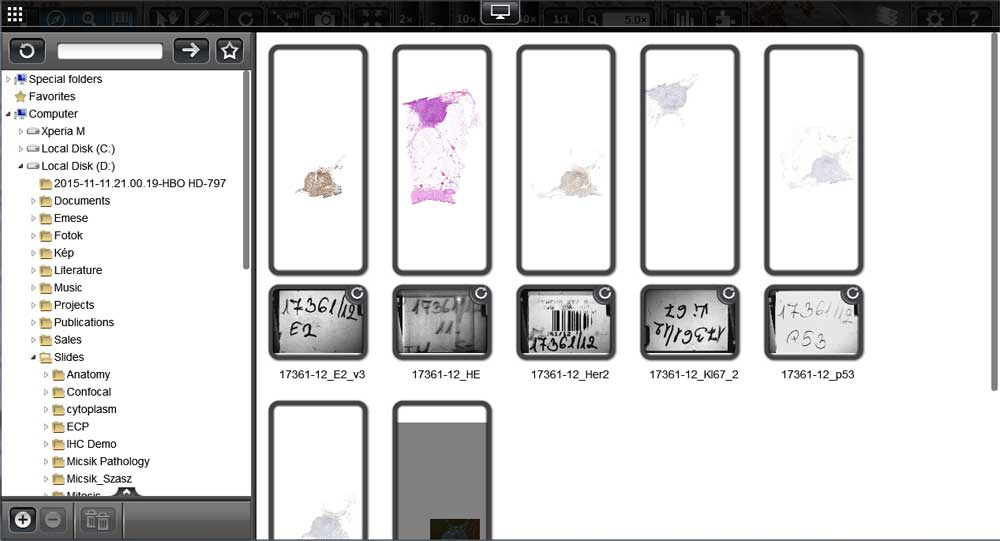
- Case preview: A slide preview panel with all slides within a case is integrated into the main screen.
- Slide stack visualization: Slides of serial sections with different stainings can be organized and then aligned in a slide stack.
Advanced file management
SlideViewer opens several 3rd party digital slides: SVS files (Aperio by Leica Biosystems) and NDP, VMS, VMU files (Hamamatsu) and supports multiple file export formats: DICOM (optional), TIFF, JPEG, SVS, MRXS.
Key Features for Research Microscopy
- Tracking history is available in high resolution recalling the history of reviewed regions of the slide step by step.
- Autofluorescence compensation: FFPE (formalin-fixed paraffin-embedded) samples typically contain fluorescent background signals because of the autofluorescent characteristic of formalin. The image compensation feature limits the visual appearance of the fluorescent stain in the background and highlights the fluorescent signals of the relevant protein.
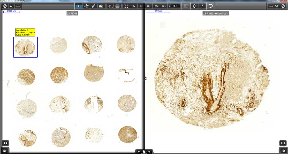
- TMA annotations: A new annotation can be defined and maintained by SlideViewer for TMA cores as well. The new TMA annotation is available in different sizes and colors. It also supports the attachment of remarks to graphical TMA annotations.
- Supports SlideDriver for microscope-like navigation on digital slides.
- Scoring guides: A new grading and scoring guideline table is available in order to support the diagnostic process.
- Native TIFF export: The annotations, the current view image and the whole slide image can be exported as a high-resolution TIFF image.
- Histogram shows the representation of the distribution of colors in a digital slide. This function helps optimize digital slides based on black, white and gamma values. The new histogram supports 16-bit image presentation.
- Z-stack prefetch supports seamless transition of the layers and optical sections in confocal or Z-stack slides.
Available Plugins
- DDIC (Digital Differential Interference Contrast) is a contrast enhancement function to reveal small details of the tissue (Windows and Mac)
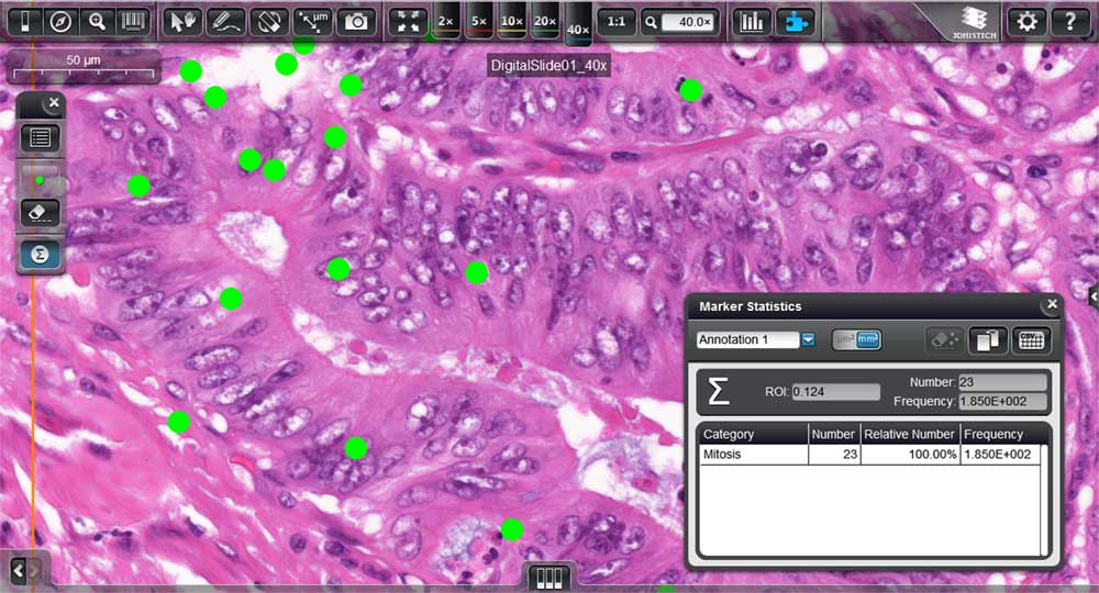
- Marker Counter is a software module for counting stained cells, immune-positive cell structures or mitosis in the tissue (Windows only)
- DICOM Snapshot: The snapshot export function is now available in the DICOM image format as well
- 3DPeek, a plugin for optimum Z-stack 3D visualization: This function provides a window into the investigation of histological structures in real 3D view – optimal for brightfield, fluorescent and also for confocal slide visualization (Windows only)
- Gradient Map Visualization (Windows and Mac)
- TMA Spot Detection* (Windows only)
- QuantCenter* image analysis modules for automated quantitative evaluation (starting with QuantCenter 2.3) (Windows only)
*Note that these functions/plugins are license dependent and therefore not free.
Other Functions
- Slide Upload: The improved slide upload makes it possible to upload slides or folders (containing slides) at a much faster (approx. 10x) speed than before.
- Slide anonymity: By using this feature of the Slide Converter, the label area image can be removed from the slide. The anonymized slide can then be shared freely and used for teaching and publishing purposes.
- Zoom limit Setting: In the Settings menu the default zoom limit can be increased in order to make digital zooming available for higher magnification.
- Tooltip: Detailed function description appears automatically by navigating the mouse arrow above any function button.
- Multi-instance enables the operating system to run multiple SlideViewer applications simultaneously on the same computer.
- Multi-core support: SlideViewer supports high performance multi-core processor operation. The number of processor cores used for running the SlideViewer application faster and efficiently can be set in the Settings menu.
Please Note
- SlideViewer can be installed and used in parallel with ClinicalViewer on the same computer.
- As SlideViewer 2.5 is the direct successor of CaseViewer 2.4, installing SlideViewer on a computer will overwrite any CaseViewer version.
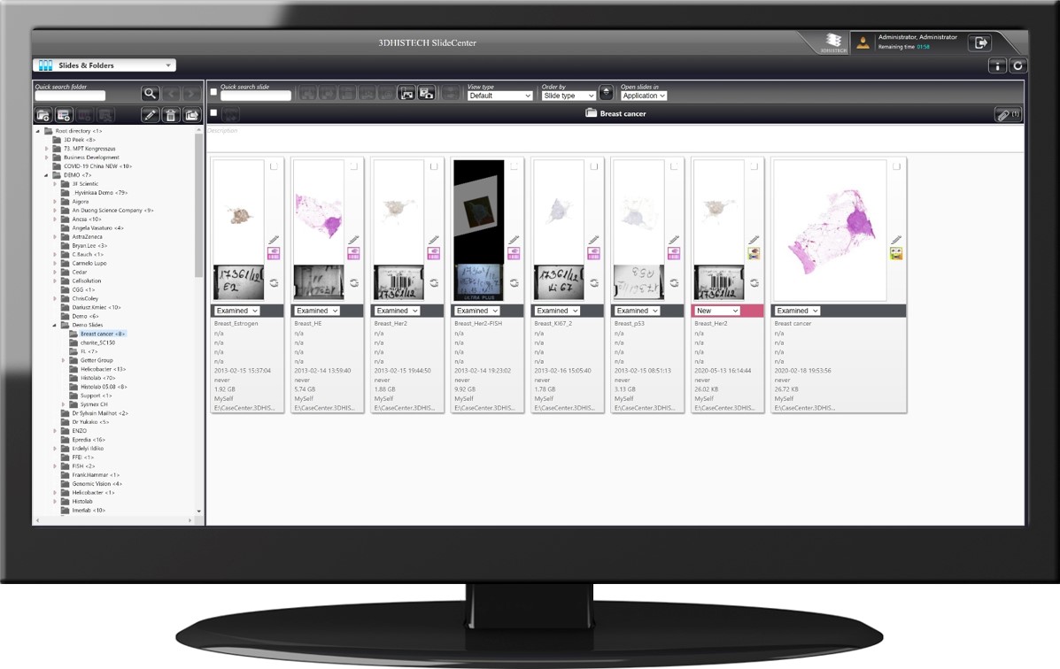
- SlideViewer will continue to be fully compatible with both CaseCenter and SlideCenter.
- License-dependent (paid) TMA plugins will continue to work on SlideViewer.
- New SlideViewer is fully compatible with new QuantCenter version 2.3. Note however that previous QuantCenter versions will not be compatible with SlideViewer version 2.5 – users will need to use CaseViewer 2.4 or earlier.
Please visit this page to read the SlideViewer FAQ.
Download SlideViewer 2.8 for Windows
Download CaseViewer 2.2.1 for Macintosh (only with CaseCenter/SlideCenter)
Please note that the SlideViewer installer now also includes the Slide Converter for TIFF and other third-party slide exports.
Note: the username and password registered on the 3DHISTECH website cannot be used to log in to the SlideCenter server.