• Skip to primary navigation
  • Skip to main content

The Digital Pathology Company

  • ‹ Company website
  • For Partner
  • Software Downloads
  • Old navigation ›
    • Homepage
    • Solutions
      • Solutions for Diagnostics
      • Solutions for Research
      • Solutions for Education
      • Solutions for Veterinary Pathology
      • Solutions for Geology
      • Scanning Service
    • Diagnostics
      • PANNORAMIC Pathology Management System
        • CaseManager DX
          • ClinicalViewer
        • DiagnosticApplications
      • PANNORAMIC Diagnostic Scanners
        • PANNORAMIC Flash DESK DX
        • Digital Pathology Solution with The Pannoramic 75 DX Digital Scanner
        • Digital Pathology Solution with The Pannoramic 150 DX Digital Scanner
        • PANNORAMIC 250 Flash III DX
        • PANNORAMIC™ 480 DX
        • PANNORAMIC 1000 DX
        • Legacy Scanners
      • Laboratory Information System
        • Track & Sign
        • CentralLogService Application (CLS) – Cybersecurity
      • Laboratory Components
        • MacroStation
        • SlideDriver
    • Research
      • SlideManager
      • PANNORAMIC™ Digital Slide Scanners
        • PANNORAMIC 1000
        • PANNORAMIC™ 480
        • PANNORAMIC 250 Flash III
        • PANNORAMIC Confocal
        • PANNORAMIC SCAN II
        • PANNORAMIC MIDI II
        • Pannoramic™ MIDI III
        • PANNORAMIC DESK II DW
      • Tissue Microarrayers
        • TMA Grand Master
        • TMA molmed
        • TMA Master II
        • TMA Applications
          • TMA Control
          • TMA Module
      • PANNORAMIC-X
      • Digital Microscopes – Viewers
        • SlideViewer
        • WebViewer
        • InstantViewer
        • iPad Viewer
        • 3DView
      • Image Analysis – QuantCenter
      • Slide Management – SlideCenter
        • SlideMaster
      • SimpleSlideInterface
      • ESchool
      • Software Downloads
    • Sales
      • Newsletter
      • Inquiry
      • Distributor Network
      • Become a Distributor
    • Support
      • Technical Support Form
      • Activity Report Form for Partners
      • Knowledge Base
        • Reference Institutes
        • Publications
          • Publications
          • TMA Grand Master publication list
          • TMA Master publication list
        • Case Studies
        • Whitepapers
        • Tutorials
      • Request Demo
    • News & Events
      • Events
      • News
    • About Us
      • Awards
      • Quality Management System
      • Funds
      • Funds (HU)
      • Contact Us
    • Feedback
  • Partner Home
  • Account
  • Log Out
Home / Research / QuantCenter and Applications

QuantCenter and Applications

QuantCenter is a multiple-module image analysis platform designed for whole-slide quantification in histopathology and molecular pathology. Thanks to its wide range of linkable modules and the ability to create their own image analysis algorithm, QuantCenter provides researchers with maximum flexibility in quantitative image analysis, contributing to world-class research results.

Individual applications created in QuantCenter can also be uploaded to SlideManager, for convenient execution on the research study samples.

quantcenter modules

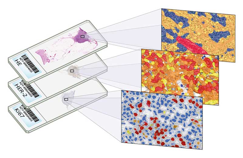
Quantification Modules

QuantCenter provides a range of image analysis modules that can be freely combined for best results.

HistoQuant

HistoQuant is an image segmentation module which identifies stained tissue elements based on color and intensity features.

PatternQuant

PatternQuant is a trainable pattern recognition module for tissue classification, tissue pre-segmentation and identification of several tissue structures.

PatternQuantPlus

PatternQuant Plus is an improved alternative to PatternQuant for more challenging samples and for complex tissue segmentation projects when thorough training using deep learning on multiple slides is needed.

NuclearQuant

NuclearQuant is designed for cell nuclei detection and quantification of IHC stained samples.

MembraneQuant

MembraneQuant is a cell membrane detection software application that can be used for the quantification of IHC stained histological samples.

CellQuant

The Swiss army knife of 3DHISTECH’s image analysis tools, CellQuant is a versatile cell detection application, an optimal solution for investigating various IHC stainings (best suited for Ki67 slides).

DensitoQuant

DensitoQuant is an easy-to-use, fast and accurate, stain-intensity-based IHC quantification tool.

FISHQuant

FISHQuant is a powerful cancer and cytogenetic application dedicated to quantifying  FISH  (Fluorescent In  Situ  Hybridization) signals on tissue samples of solid tumour diseases like breast cancer, lung cancer, sarcoma symptoms, and lymphomas.

CISHQuant

CISHQuant quantifies CISH (Chromogenic In Situ Hybridization) stained samples.

ScriptQuant

ScriptQuant provides a shell for user-created image analysis scripts in Python for maximum flexibility for the research user, enabling them to run their own image analysis algorithms within the QuantCenter framework.

SimpleSlideInterface

SimpleSlideInterface enables easy access to slide-related data, allowing you to develop your own application.

Quantification Tools

QuantCenter also provides assistance for studies performed on multiple slides, quantification processes can be performed both on the client and server side, and their results can be examined with data mining tools.

Server-side Analysis – QuantServer

QuantServer module allows you to execute processing and store the analyzation results on a dedicated server.

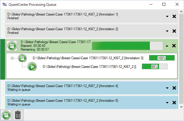
Batch Analysis – Processing Queue

Using the batch analysis module, multiple digital slides can be examined in the background on your computer, saving a significant amount of time.

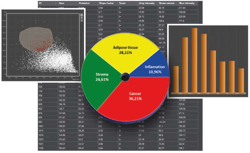
Data Visualization – DVT

An integrated data visualization mode is available for each module. The result can be visualized on a  table,  scatterplot, pie chart or a histogram by using this feature.

Quantification Examples

The gallery below contains some examples of how widely our quantification modules can be used.

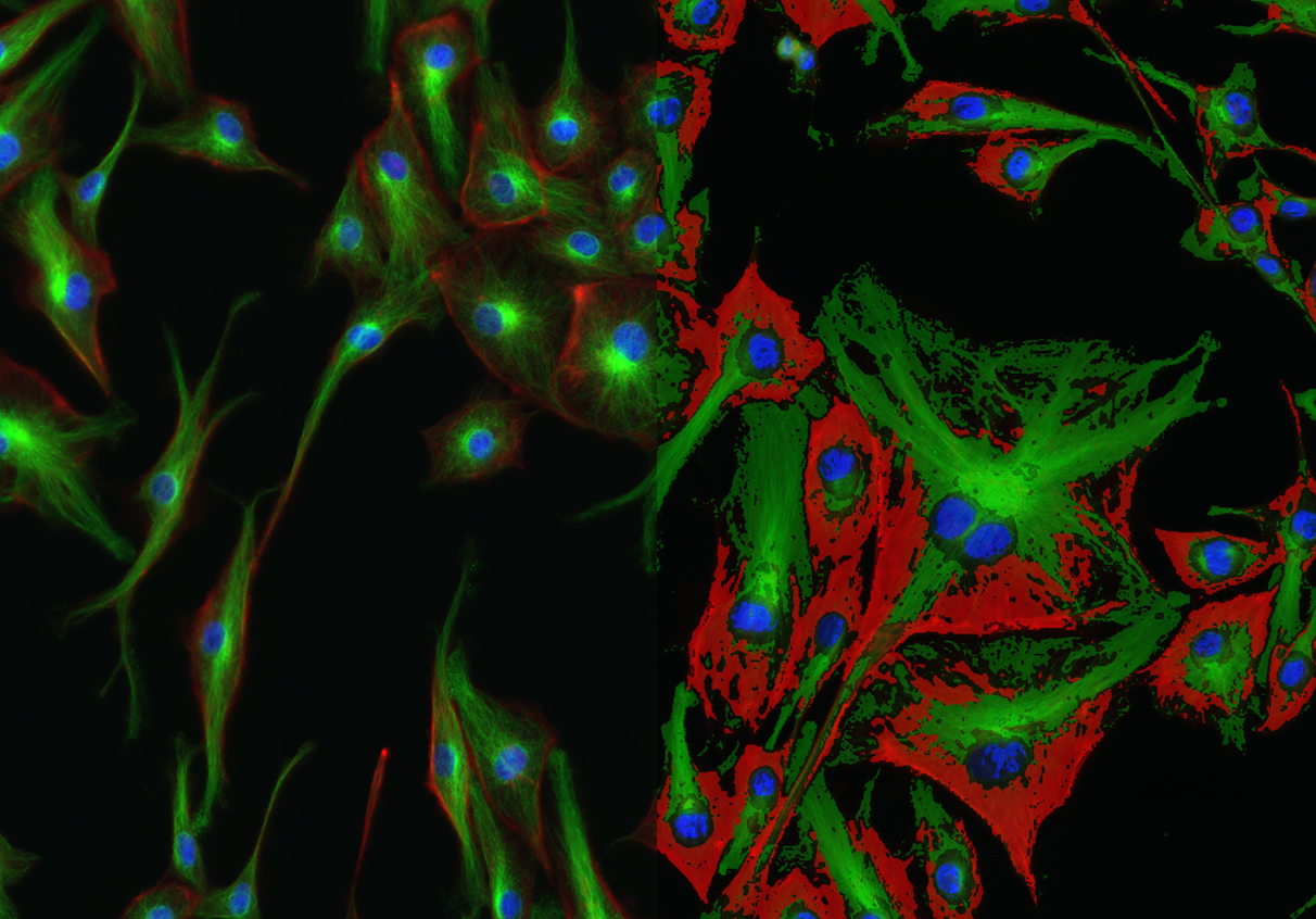
Bovine pulmonary artery endothelial cell quantification by HistoQuant

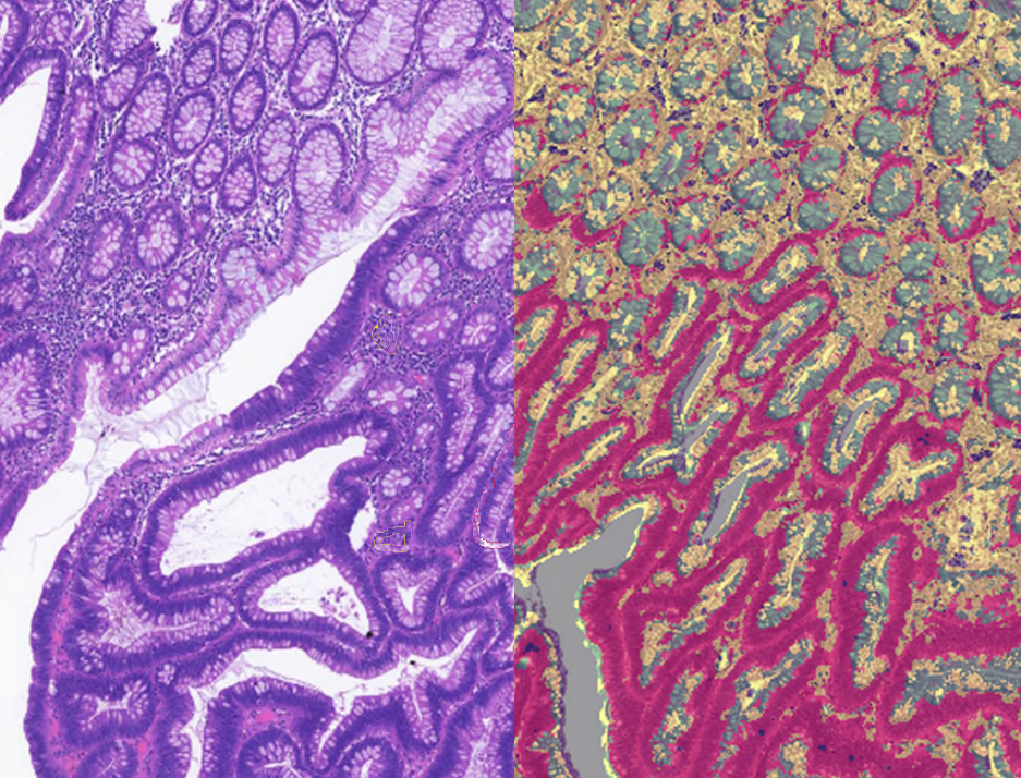
Cyprinus carpio epithelium analysis by PatternQuant

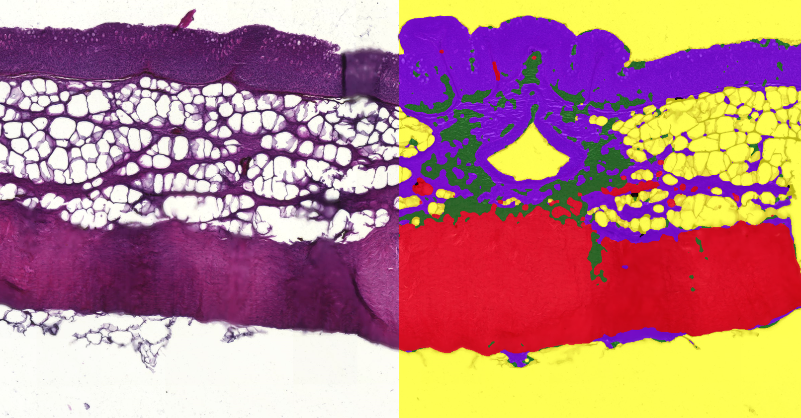
Elongated villous structure with multi-layered nuclei analysis by PatternQuant

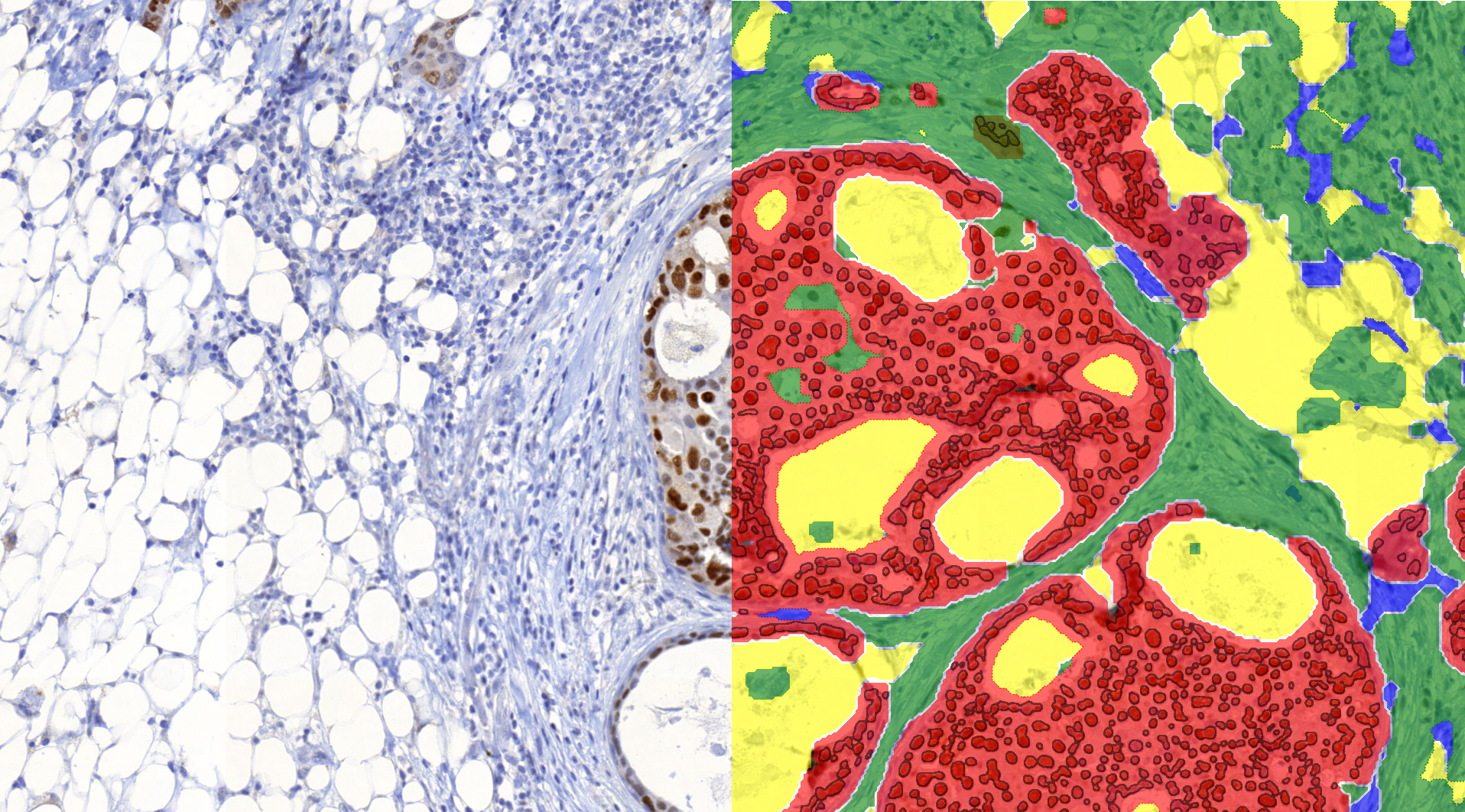
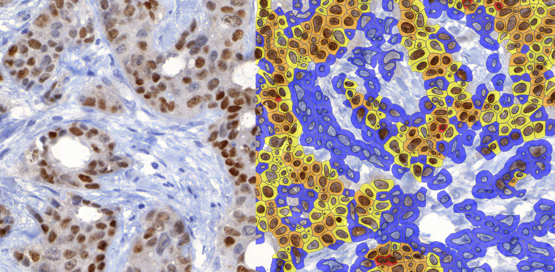
Estrogen stained infiltrating ductal carcinoma in breast tissue by PatternQuant and NuclearQuant

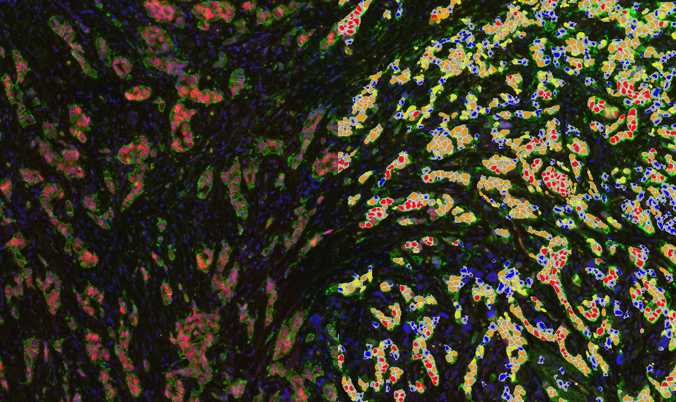
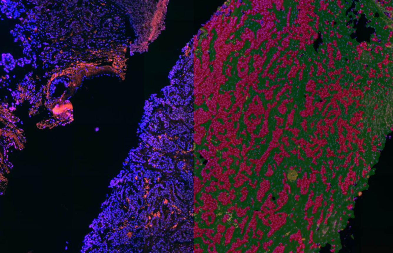
Estrogen-Her2 stained breast tissue analysis by CellQuant

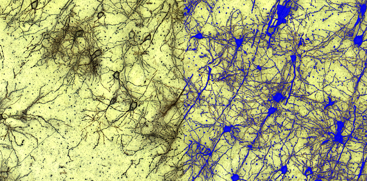
Golgi stained neuron network detection in the rat cortex by HistoQuant

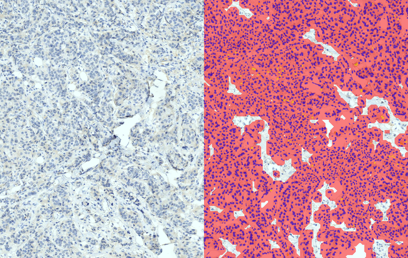
Nuclei classification in the tumour area of progresterone stained breast tissue by NuclearQuant

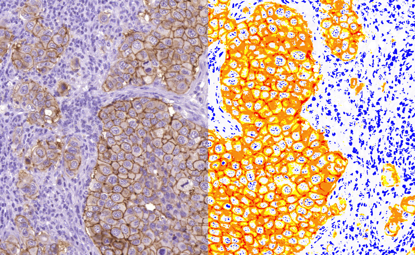
Her2 stained breast tissue analysis by DensitoQuant

Her2-Cep17 DNA stained breast tissue classification by HistoQuant

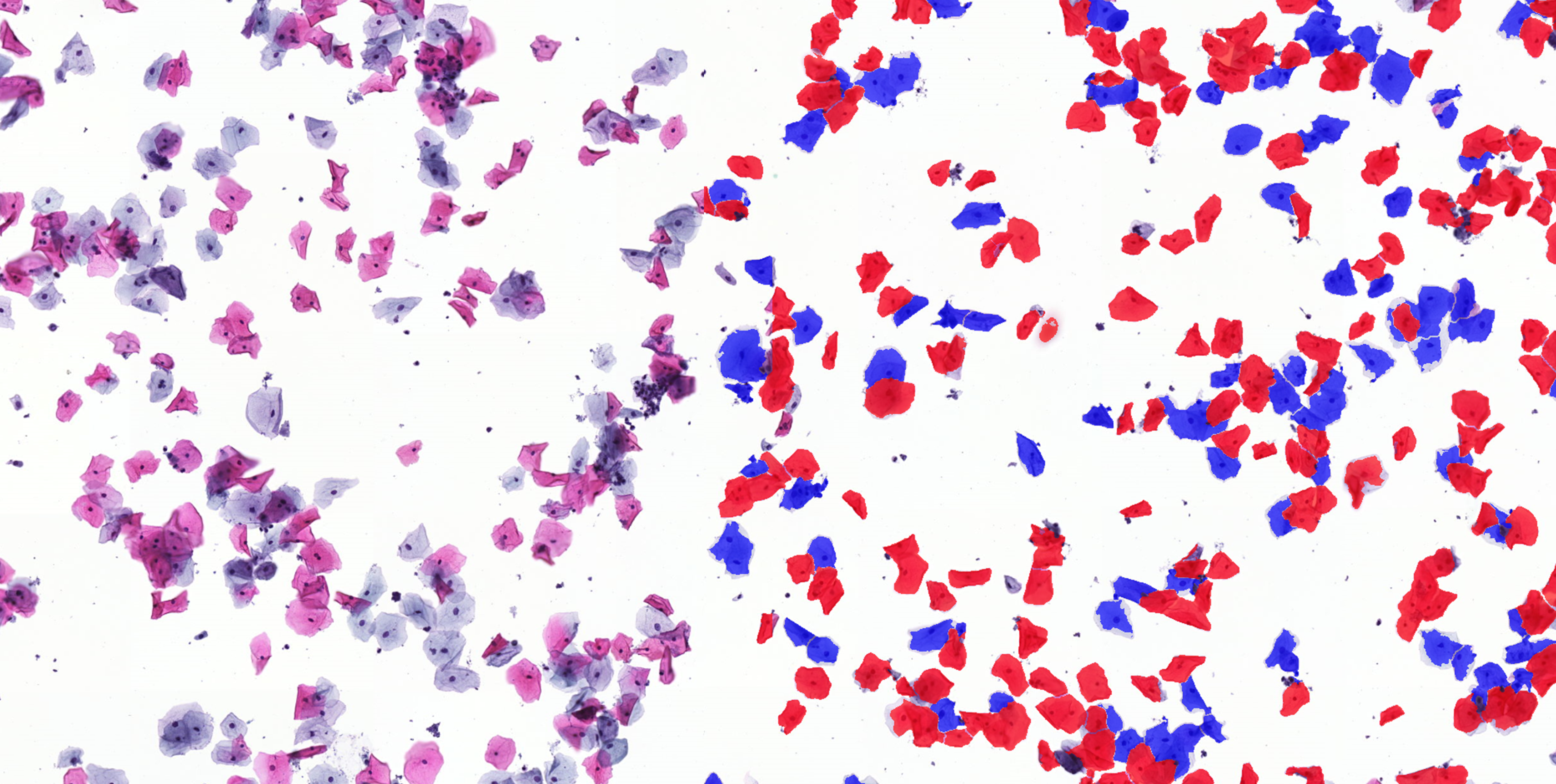
Nuclei classification on cytology smear sample by HistoQuant

Progresterone stained cytoplasm classification by CellQuant

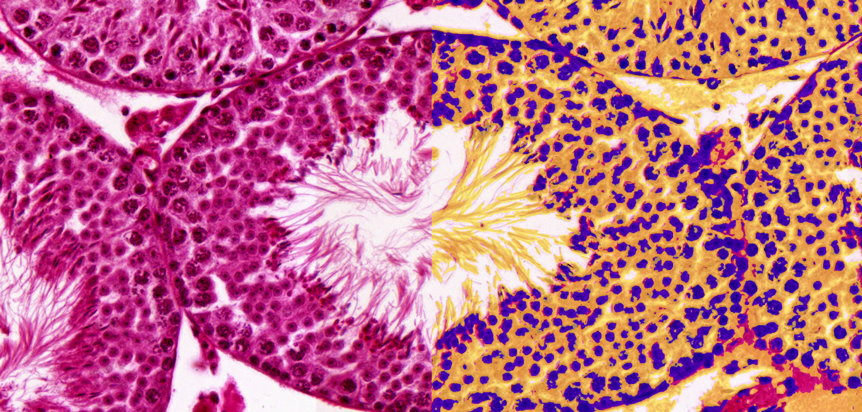
Testicle organ with sperm cells analysis by HistoQuant

  • ‹ Company website
  • For Partners
  • Software Downloads
  • facebook
  • X
  • linkedin
  • youtube
  • instagram

Partners

Login

© 2005-2025, 3DHISTECH Ltd. All rights reserved.
H-1141 Budapest, Öv u. 3., Hungary

We are using cookies to give you the best experience on our website.

You can find out more about which cookies we are using or switch them off in .

3DHISTECH Ltd.
Powered by  GDPR Cookie Compliance
Privacy Overview

This website uses cookies so that we can provide you with the best user experience possible. Cookie information is stored in your browser and performs functions such as recognising you when you return to our website and helping our team to understand which sections of the website you find most interesting and useful.

Strictly Necessary Cookies

Strictly Necessary Cookie should be enabled at all times so that we can save your preferences for cookie settings.

If you disable this cookie, we will not be able to save your preferences. This means that every time you visit this website you will need to enable or disable cookies again.

Google Analytics

This website uses Google Analytics to collect anonymous information such as the number of visitors to the site, and the most popular pages.

Keeping this cookie enabled helps us to improve our website.

Please enable Strictly Necessary Cookies first so that we can save your preferences!