• Skip to primary navigation
  • Skip to main content

The Digital Pathology Company

  • ‹ Company website
  • For Partner
  • Software Downloads
  • Old navigation ›
    • Homepage
    • Solutions
      • Solutions for Diagnostics
      • Solutions for Research
      • Solutions for Education
      • Solutions for Veterinary Pathology
      • Solutions for Geology
      • Scanning Service
    • Diagnostics
      • PANNORAMIC Pathology Management System
        • CaseManager DX
          • ClinicalViewer
        • DiagnosticApplications
      • PANNORAMIC Diagnostic Scanners
        • PANNORAMIC Flash DESK DX
        • Digital Pathology Solution with The Pannoramic 75 DX Digital Scanner
        • Digital Pathology Solution with The Pannoramic 150 DX Digital Scanner
        • PANNORAMIC 250 Flash III DX
        • PANNORAMIC™ 480 DX
        • PANNORAMIC 1000 DX
        • Legacy Scanners
      • Laboratory Information System
        • Track & Sign
        • CentralLogService Application (CLS) – Cybersecurity
      • Laboratory Components
        • MacroStation
        • SlideDriver
    • Research
      • SlideManager
      • PANNORAMIC™ Digital Slide Scanners
        • PANNORAMIC 1000
        • PANNORAMIC™ 480
        • PANNORAMIC 250 Flash III
        • PANNORAMIC Confocal
        • PANNORAMIC SCAN II
        • PANNORAMIC MIDI II
        • Pannoramic™ MIDI III
        • PANNORAMIC DESK II DW
      • Tissue Microarrayers
        • TMA Grand Master
        • TMA molmed
        • TMA Master II
        • TMA Applications
          • TMA Control
          • TMA Module
      • PANNORAMIC-X
      • Digital Microscopes – Viewers
        • SlideViewer
        • WebViewer
        • InstantViewer
        • iPad Viewer
        • 3DView
      • Image Analysis – QuantCenter
      • Slide Management – SlideCenter
        • SlideMaster
      • SimpleSlideInterface
      • ESchool
      • Software Downloads
    • Sales
      • Newsletter
      • Inquiry
      • Distributor Network
      • Become a Distributor
    • Support
      • Technical Support Form
      • Activity Report Form for Partners
      • Knowledge Base
        • Reference Institutes
        • Publications
          • Publications
          • TMA Grand Master publication list
          • TMA Master publication list
        • Case Studies
        • Whitepapers
        • Tutorials
      • Request Demo
    • News & Events
      • Events
      • News
    • About Us
      • Awards
      • Quality Management System
      • Funds
      • Funds (HU)
      • Contact Us
    • Feedback
  • Partner Home
  • Account
  • Log Out
Home / Research / Tissue Microarrayers / TMA Module

TMA Module

TMA Module is an application that enables researchers to deal with a large number of samples very easily in a structured way and makes the evaluation and scoring of several hundred samples possible.

Tissue microarrays represent a significant move towards economy and constant quality in immuno-histochemistry: Up to a couple hundreds of samples can be fitted on a single slide and all these samples are stained at the same time. Little arrays are taken out of the specimen with a needle-like tool and then aligned in blocks. After sectioning these blocks are put on a slide and stained homogeneously.

TMA module

TMA is useful when the researcher is only looking for signs of specific parts (proteins usually) in the sample. With the means of immuno-histochemistry it is possible to detect and to visibly differentiate the desired proteins which means it can be effectively used e.g. in cancer diagnosis. It is a very rational step to fit as many samples (here: cores) on one slide as possible because staining in constant quality is essential in immunohistochemistry. However, this creates a problem traditional microscopy cannot cope with efficiently.

The much greater amount of samples requires much greater administrative work. You have to find the core, record your score and then move to the next position. It is easy to miss. If you want to compare your previous scores with your present ones it is almost impossible because navigation in the traditional microscope was not invented for this kind of job.

This is one of the fields where virtual microscopy clearly shows its strengths. By using the TMA Module software, you can enjoy all the benefits of tissue microarrays.

Main characteristics

  • Project-based workflow: link multiple blocks of multiple slides together in one project. This enables you to deal with huge number of samples very easily in a structured way. Slides can be added to and removed from an existing project.
  • Data of samples and blocks and also the map of blocks are presented in an .xls file.
  • Based on the map of blocks the program detects the cores and assigns the selected attributes to them.
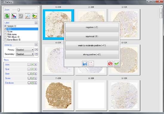
  • Scoring of cores is easy and convenient: TMA is integrated with SlideViewer (former CaseViewer) so seamless zooming and navigation is available. Range of values of scoring can be set by the user. After scoring the virtual objective jumps to the next core so you don’t have to find it. The scoring results are saved in the XLS database.
  • Simple (1-dimensional) and combined (2-dimensional) scoring schemes can be set by the user.
  • The cores can be filtered based on scores and other attributes, including measurement data created by the quantification software applications.
  • The data of scoring can be analyzed with third-party software.

Product brochure

Would you like to know more about our solutions? Please get in touch!

Related Products

TMA Master II

TMA Master II is the smallest fully automated tissue microarrayer on the market, it easily fits any laboratory bench.

TMA Control Software

The TMA Control software is the perfect solution for TMA block design and creation.
TMA Control software by 3DHISTECH

TMA Grand Master

The TMA Grand Master is the fastest, highest capacity and most user friendly tissue microarrayer on the market.

  • ‹ Company website
  • For Partners
  • Software Downloads
  • facebook
  • X
  • linkedin
  • youtube
  • instagram

Partners

Login

© 2005-2025, 3DHISTECH Ltd. All rights reserved.
H-1141 Budapest, Öv u. 3., Hungary

We are using cookies to give you the best experience on our website.

You can find out more about which cookies we are using or switch them off in .

3DHISTECH Ltd.
Powered by  GDPR Cookie Compliance
Privacy Overview

This website uses cookies so that we can provide you with the best user experience possible. Cookie information is stored in your browser and performs functions such as recognising you when you return to our website and helping our team to understand which sections of the website you find most interesting and useful.

Strictly Necessary Cookies

Strictly Necessary Cookie should be enabled at all times so that we can save your preferences for cookie settings.

If you disable this cookie, we will not be able to save your preferences. This means that every time you visit this website you will need to enable or disable cookies again.

Google Analytics

This website uses Google Analytics to collect anonymous information such as the number of visitors to the site, and the most popular pages.

Keeping this cookie enabled helps us to improve our website.

Please enable Strictly Necessary Cookies first so that we can save your preferences!